
Quantifies tumor content and generates tumor density heatmap to guide ROI selection for macrodissection in colorectal cancer
Vendor
Indica Labs
Company Website
Overview
CRC Macrodissect AI is an AI-powered tool that quantifies tumor content and guides ROI selection to enhance macrodissection workflows and downstream molecular analysis in colorectal carcinoma.
Intended Use
Research Use Only
Inputs
H&E whole slide images from primary and metastatic CRC resections, excisions, and/or core needle biopsies.
Key Outputs
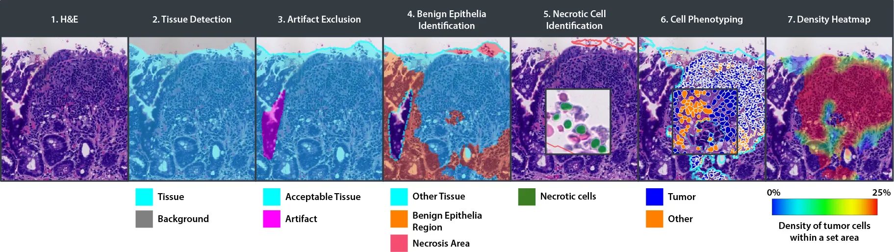
- Tissue Detection
- Artifact Exclusion
- Benign Epithelial Identification
- Necrotic Cell Identification
- Cell Phenotyping
- Density Heatmap
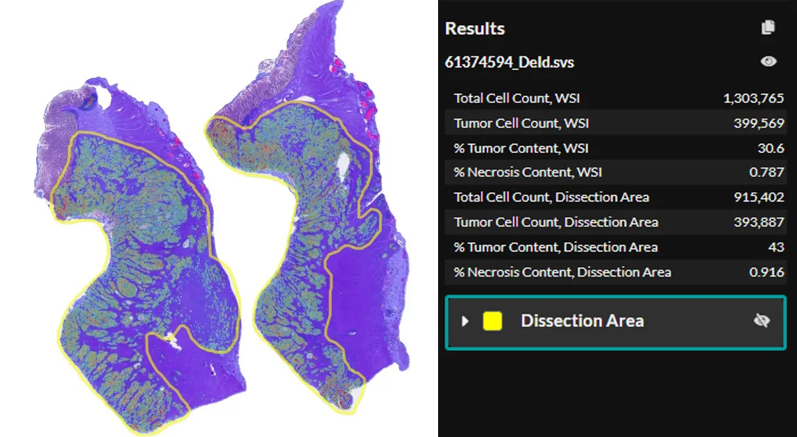
- Percent tumor content for whole slide image and dissection ROIs
File Formats:
- Non-proprietary (JPG, TIF, OME.TIFF, DICOM [DCM*])
- Leica (SVS, AFI, SCN, LIF)
- Hamamatsu (NDPI, NDPIS)
- Philips (iSyntax, i2Syntax)
- 3DHistech (MRXS)
- Nikon (ND2)
- Akoya (QPTIFF, component TIFF)
- Olympus / Evident (VSI)
- Zeiss (CZI)
- Ventana (BIF)
- KFBIO (KFB, KFBF)
- *whole-slide images
Benefits
Confidence in Results
CRC Macrodissect AI reliably quantifies tumor content for downstream molecular analysis, ensuring the quality of downstream test results.
Streamline Workflows and Save Resources
With automated tumor content analysis, you can streamline your ROI selection process and save time.
Auditable Process
Create an auditable macrodissection workflow, ensuring transparency, efficiency, and accuracy of molecular test results.
Other
CRC Macrodissect AI bridges the gap between anatomic and molecular pathology by simplifying the macrodissection process. Pathologists need only use the intuitive annotation tools provided in HALO AP® to select areas of high tumor density for downstream analysis by following the easy-to-read heatmap. Tumor content results for annotated ROIs are updated in real time.
Macrodissection Reinvented
CRC Macrodissect AI enhances macrodissection workflows with precision and automation. H&E slides are scanned into HALO AP®, where CRC Macrodissect AI detects all tissue present on the slide, removing background glass and artifacts from the analysis. Benign epithelial and regions of necrosis are classified separately, and their cell count is added to the tumor content results. Cells are then phenotyped as either ‘tumor’ or ‘other’ cells. A detailed tumor density heatmap is generated, which assists pathologists in creating precise ROI annotations for downstream macrodissection.