
Quantifies HER2, ER, PR, and Ki67 IHC expression levels in primary invasive breast cancer
Vendor
Indica Labs
Company Website
Overview
Breast IHC AI is an AI-powered image analysis tool that accurately detects regions of invasive tumor, standardizes scoring of HER2, ER, PR,and Ki67, and enables detection of low and ultralow HER2 expression in breast cancer.
Intended Use
Breast IHC AI is For Research Use Only and not intended for clinical diagnostic use.
Inputs
WSI of resections, excisions, and/or biopsies from primary invasive breast cancer
Key Outputs
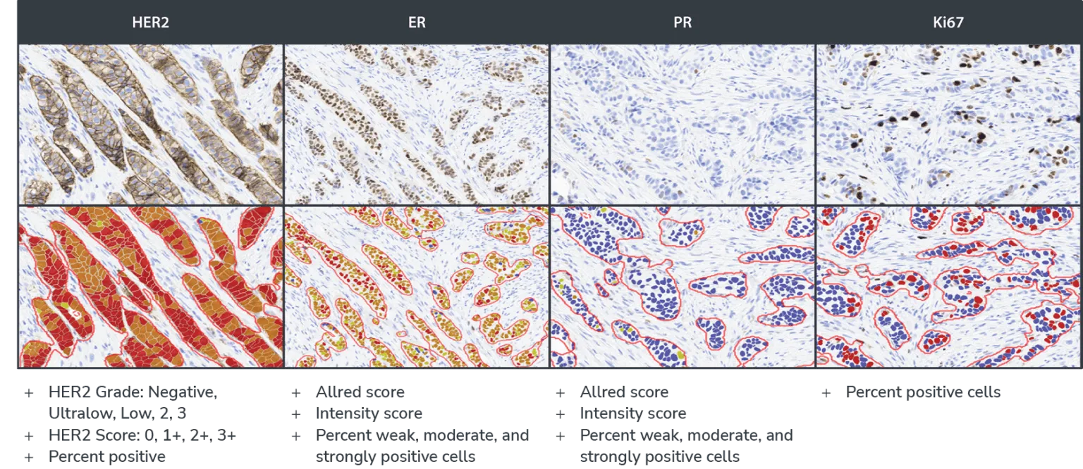
- HER2: HER2 Grade: Negative, Ultralow, Low, 2, 3; HER2 Score: 0, 1+, 2+, 3+; Percent positive
- ER and PR: Intensity Score: percent of weak, moderate, and strong positive cells; AllRed Score
- Ki67: Percent of positive cells
Supported Clones
- HER2: 4B5; CB11
- ER: EP1; SP1
- PR: PgR636; Pgr A/B: 16+SAN27
- Ki67: MIB1, SP6
File Formats
- Non-proprietary (JPG, TIF, OME. TIFF, DICOM [DCM*])
- Leica (SVS, AFI, SCN, LIF)
- Hamamatsu (NDPI, NDPIS)
- Philips (iSyntax, i2Syntax)
- 3DHistech (MRXS)
- Nikon (ND2)
- Akoya (QPTIFF, component TIFF)
- Olympus / Evident (VSI)
- Zeiss (CZI)
- Ventana (BIF)
- KFBIO (KFB, KFBF)
- *whole slide images
Benefits
Standardize Biomarker Evaluation
Breast IHC AI standardizes biomarker scoring and reduces interobserver variability in the IHC evaluation process, without compromising accuracy.
More Efficient Workflow
Automated biomarker evaluation reduces the workload for pathologists and researchers while delivering accurate and efficient results.
Complement Your Expertise
Breast IHC AI provides consistent, standardized measurements so you are free to apply your expertise where it’s needed most, in the interpretation of results to make informed decisions.
Comprehensive Analysis
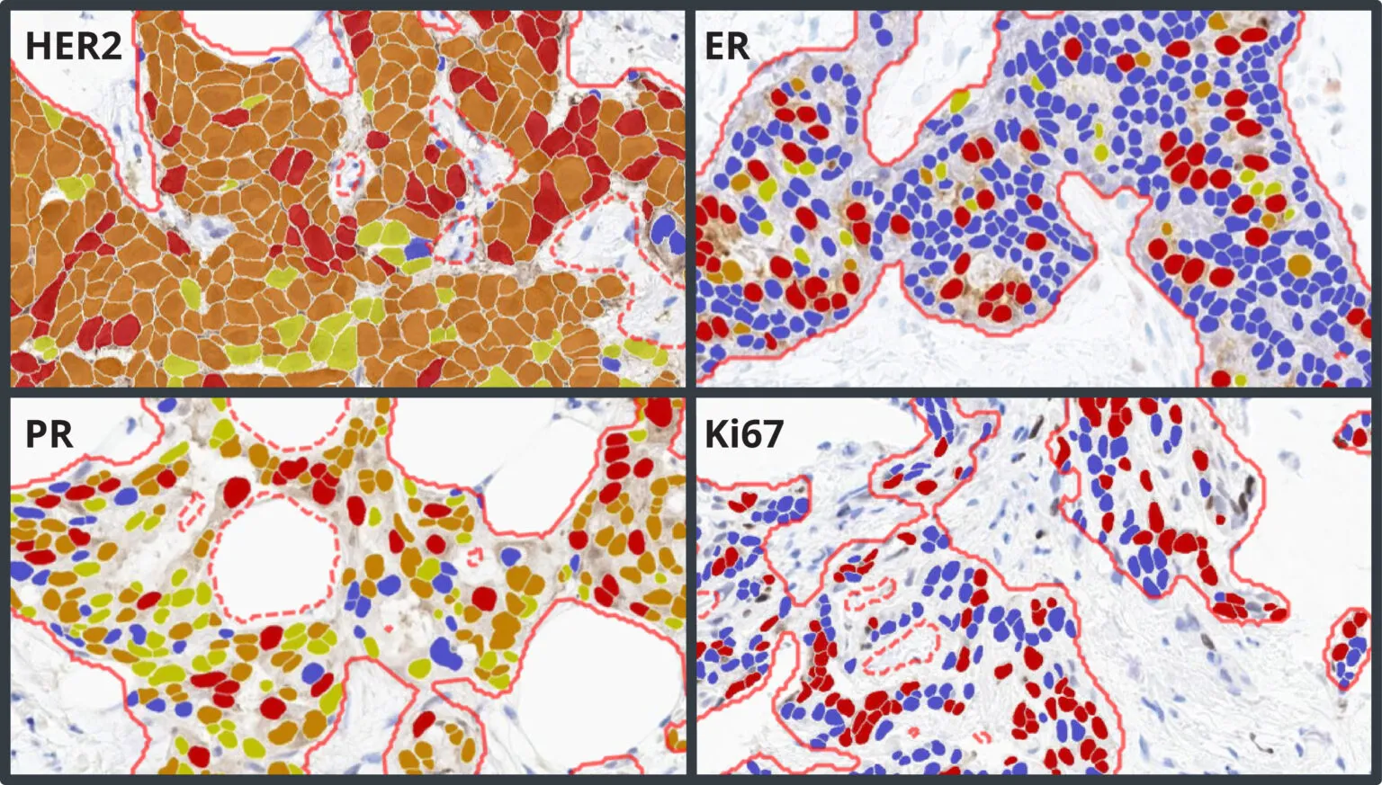
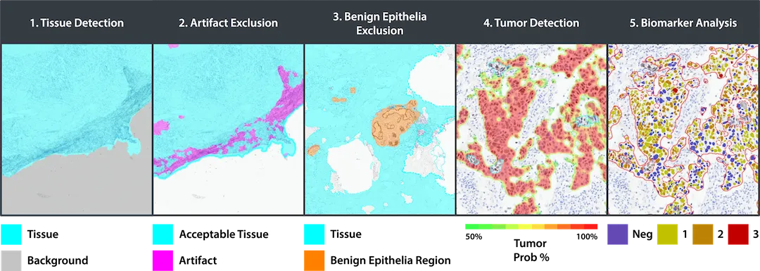
Breast IHC AI includes AI-based analysis assays for automated scoring of HER2, ER, PR, and Ki67, including analysis of HER2 low and ultralow expression. Each assay has built-in artifact exclusion, benign epithelial region exclusion, and tumor detection steps to ensure that biomarker analysis is performed accurately and consistently each and every time. Tumor cells are analyzed for expression of HER2, ER, PR, and Ki67, and a comprehensive set of results and markups are generated for each image, including clinical score and percentage positivity.
Accurately Detect HER2 Low and Ultralow Expression in Breast Cancer
The HER2 algorithm detects all levels of expression, including low and ultralow levels and outputs a Grade of Negative, Ultralow, Low, Grade 2, and Grade 3 with a corresponding Score of HER2 0, 1+, 2+, and 3+. The software assists the pathologist by automatically reporting the percentage of positive cells and biomarker scores at the slide level, along with image analysis masks which can be viewed in the HALO AP® platform.
Seamless Deployment in HALO AP®
Breast IHC AI is deployed and fully integrated into HALO AP®, the AI-powered, pathologist-driven platform for anatomic pathology workflows from Indica Labs.